Eye Simulator
An eye simulator is a computer simulator that is used across healthcare simulation to educate optometrists and ophthalmologists to assess, diagnose and manage vision changes and eye care. Eye simulators combine computers, external or internal images of the eye, and a simulated ophthalmoscope. They can enhance education for optometrists and ophthalmologists by teaching them to recognize common eye problems. Instructors can provide guidance and feedback in real-time, which also enhances education.
Virtual patient simulators for ophthalmology have been well-tolerated by learners and have been validated for the assessment of clinical competencies for surgical certification. Eye simulators are different from eye models, which are three-dimensional models used to study the anatomy of the eye. Eye models are enlarged to show the parts of the eye, and usually come apart to display external and internal structures.
Learning to properly use an ophthalmoscope benefits several health care disciplines, including nurse practitioners, physicians, physician assistants, optometrists and ophthalmologists. However, the latter two disciplines in particular benefit from the adoption and use of eye simulators with advanced technology (as described below).
Sponsored Content:
Optometrists provide primary vision care, which ranges from sight testing and correction to the diagnosis, treatment and management of vision changes. Optometry involves performing eye exams and vision tests, prescribing and dispensing corrective lenses (such as eyeglasses and contact lenses), detecting certain types of eye abnormalities and prescribing medications for certain eye diseases.
Ophthalmologists are medical doctors who specialize in eye and vision care, and are licensed to practice medicine and perform surgery. An ophthalmologist diagnoses and treats all eye diseases, performs eye surgery, and prescribes and fits corrective lenses to correct vision problems.
Types of Eye Simulators
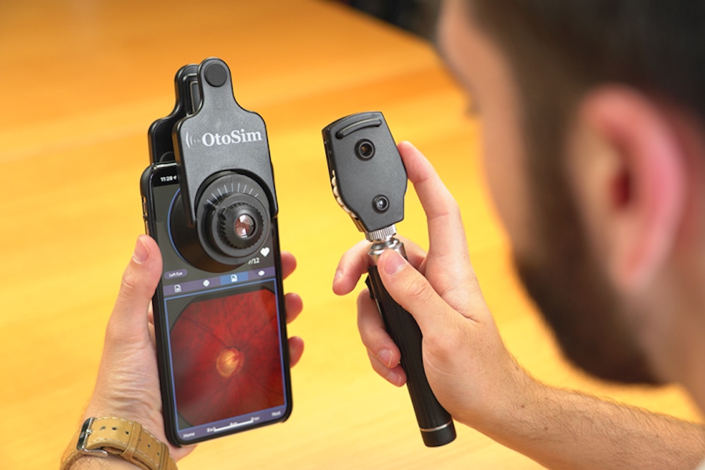
OtoSim: OtoSim offers two simulation products for ophthalmological skills training, the OphthoSim and OphthoSim Mobile. Both provide learners with the opportunity to diagnose and treat eye pathologies using an immersive learning platform. The OphthoSim includes a desktop model base and simulated ophthalmoscope, while the mobile version includes a silicone eyeform that clips to a smartphone and a simulation ophthalmoscope. Both products also come with a content library of over 200 high-resolution retinal images, training and testing modules, and an instructor portal.
Sponsored Content:
The ophthalmoscope contains position-detecting hardware that enables the OphthoSim software to track the movement and orientation of the scope. Realistic eye geometry allows the iris of the eye to dilate and constrict to alter the field of view during an examination. Retina images include normal fundus, papilledema, diabetic retinopathy, glaucoma, choroidal rupture, macular degeneration, tumors and other ocular irregularities.
Eye Movement Simulator: This realistic web app simulates the movement of the external ocular muscles (EOM) and pupillary responses. It was designed in 3D, and simulates the physical examination of EOM by dragging the computer mouse as the focal point for the eyes to follow. The app can be used to recreate pathologies by making certain muscles and the corresponding cranial nerves active or inactive with simple checkboxes. The 3D-realistic rendering provides pupillary response and eye blinks.
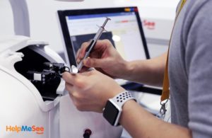
HelpMeSee Eye Surgery Simulator: A new Cataract Surgery Simulator was unveiled by the global 501(c)3 not-for-profit organization HelpMeSee, which has a mission to end the backlog of cataract blindness and visual impairment, especially in developing countries. The HelpMeSee Eye Surgery Simulator is a highly realistic virtual reality trainer for Manual Small Incision Cataract Surgery (MSICS). The system provides a scalable approach for training cataract eye surgery specialists and ophthalmologists. The overall simulator shape mimics a patient lying back, with the trainee sitting at the top of the patient’s head (superior approach). The trainee views the surgical area through a virtual microscope mimicking an ophthalmic microscope. The trainee interacts with the simulation through two haptic handpieces that mimic the grips of the surgical instruments. The trainee can adjust the height of the simulator to a comfortable position.
The HelpMeSee Eye Surgery Simulator encompasses an adaptation of an actual virtual microscope used in surgery, two haptic handpieces, a virtual syringe, the patient head and hand rest, a touchscreen user interface, powerful visuals, and simulation software, and everything required to simulate an MSICS surgery. The two handpieces and syringe represent the complete set of surgical instruments needed to perform an MSICS procedure. Programmed lessons with onscreen guides and error messages assist the student in mastering the MSICS technique and the instructor in providing objective feedback.
Eyesi Surgical is a high-end virtual reality simulator with training units that range from basic skills training to surgical procedures and management of complications. The immersive environment is achieved through the use of the OR microscope on the simulator. Learners see the virtual surgical field in stereo and high resolution while operating with lifelike surgical instruments.
The highly-realistic simulation helps teach the discrete instrument movements required to avoid undue wound stress, loss of viscoelastic, or diminished red reflex. This real-time interaction with simulated tissue increases trainees’ surgical experience without risk to real patients.
Eyesi Surgical offers training modules for cataract surgery and retinal (or posterior segment) surgery. The latter modules require a vitreoretinal interface set, which is operated just like an actual BIOM in the operating room. Feedback and evaluation are offered after each task to enable learners to systematically improve their skills.
Utilization of a BIO Simulation System
Binocular indirect ophthalmoscopy (BIO) is an essential optometric skill that students must master in order to detect sight threatening conditions such as retinal detachment and diabetic retinopathy. However, it challenging for clinical educators to provide immediate and constructive feedback to trainees. It is therefore difficult for students to improve BIO skills efficiently and effectively. The retina of a patient cannot be viewed simultaneously by student and educator, and learners are exposed to few practice patients with limited retinal conditions.
Click Here to Connect to Leading Healthcare Simulation Vendors
The Eyesi Indirect and Eyesi Direct systems (VRmagic, Mannheim, Germany) are nonsurgical retinal diagnostic simulators used to train eye care practitioners. The system hardware includes a touch-screen user interface with either a headband-mounted stereo display with two handheld fundus lenses (indirect), or a handheld direct ophthalmoscope display (direct). These systems use augmented virtual reality to incorporate virtual views of the patient face and fundus with actual views of the surrounding environment. This creates a realistic examiner experience for BIO and direct ophthalmoscopy (DO).
The software curriculum trains the mechanics of retinal examination, recognition of normal anatomic findings, and understanding of a library of pathological findings based on actual clinical patients. The software also aids in the development of the diagnostic process by incorporating diagnostic tests such as visual fields and OCT scans, and furthers the understanding of patient management through clinical questions built into each case scenario.
Eye Simulator Education Latest News

13 Medical Simulation Keywords You Need to Know About

HelpMeSee’s Healthcare Simulation Training & Partnerships Deliver New Hope for Cataract Blind

8 Medical Simulation Keywords You Need to Know About
Sponsored Content:


