Virtual Anatomy

Virtual Anatomy is a technology tool that enables interactive anatomy visualization and learning. By using this digital anatomy, learners, educators and professionals can explore and better understand the human body. Making anatomic learning virtual, the tools can encompass 3D models, animations, quizzes, augmented reality and more.
Utilizing Virtual Anatomy can help teach concepts of anatomy, physiology, muscles, the skeleton and circulatory system. The technology also span the gap between gross anatomy and state-of-the-art medical imaging. Virtual Anatomy also extends the opportunity to learn cross-sectional anatomy and radiologic imaging alongside cadaveric dissection.
Oftentimes an actual cadaver, both male and female, can be imaged and rendered into 3-D models that learners can manipulate. The learners are able to rotate these models, virtually dissect them and essentially perform the types of experiments they would normally due in an anatomy lab. For example, users can search for body parts and terms, use interactive features to change the depth of their cuts, visualize tissues through high-resolution images and obtain multiple views.
Sponsored Content:
“For early learners, there’s a lot on their plate, so the Virtual Anatomy Lab represents one more thing they need to learn and prepare for. But I think having gone through it, they now realize how well it’s prepared them to enter the clinical realm,” John R. Harrison, PhD, the director of UConn’s virtual and human anatomy labs, told the American Medical Association.
Although this tool is an effective form of advancing digital technology, Virtual Anatomy will not likely replace gross anatomy entirely. This is because many healthcare educators and leaders still believe that early exposure to real-life anatomy will become a reference point that stays with learners for the rest of their careers.
Types of Virtual Anatomy
There are many types of Virtual Anatomy learning, including the incorporation of Virtual Cadavers, Radiology (RAD) Workstations, Ultrasound sessions and volumetric reconstruction. Each type of Virtual Anatomy helps to teach skills and procedures imperative in a number of healthcare roles and positions. The information also assists in the general understanding of bodily functions, regardless of profession.
Sponsored Content:
For instance, a virtual cadaver table will display life-size virtual cadavers, which allows learners to experience gross anatomy in axial, sagittal and coronal slices. The technology also allows for full control over the clipping plane. These features further provides the ability to explore anatomical images and reinforce anatomical relationships. A virtual cadaver also provides a powerful link between cadaveric anatomy and radiology images such as CT and MRI scans.
Alternatively, Radiology (RAD) Workstations provide users with access to a library of de-identified patient images. These images include X-rays, CT scans, MRIs and diagnostic procedures using a PACS viewer. The users will then apply their anatomical knowledge to the workstation to navigate and interpret clinical and diagnostic images.
Ultrasound sessions are then used to provide learners and professions with more hands-on anatomical experiences. These sessions help users acquire knowledge of anatomical principles, surface anatomy with the location and orientation of internal anatomy. With this knowledge, the user can then perform ultrasound on patient instructors. This helps them to visualize normal anatomy in contrast with relevant pathologies using virtual sonography workstations.
Another resource, volumetric reconstruction can be used on CT and MRI scans to show learners how to use gaming technology to navigate through and explore anatomical structures from any orientation. With visualization features that enable users to quickly and effectively view and interact with MRI and CT scan data, they can practice and perform tasks in a 3D manner.
Examples of Virtual Anatomy
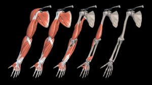
Complete Anatomy, is a cloud-based 3D Animated Learning platform from 3D4Medical that allows users to investigate the minute detail of the human anatomy in incredible 3D. Validated by an academic advisory board and in-house anatomical experts, this is one of the most accurate and complex 3D anatomical atlas platforms available today! Unlike other anatomy apps, 3D4Medical allows you to use modeling tools with 2D & 3D pens to cut, fracture, grow, spur or otherwise demonstrate pain in specific body parts, all while adding images, labels to saved 3d screens with full model interactivity.
Click here to Learn More About 3D4Medical!
Educators looking to hit the ground running can gain access to 100 full courses already developed by some of the World’s leading Subject Matter Experts! Any side by side comparison will demonstrate a higher level of model realism, from veins and arteries to system organ connectives, joints, layers and structures. 3D4Medical’s Complete Anatomy is an award-winning 3D Anatomy App available across all Apple / Android / PC devices, which provides a robust anatomical learning and teaching platform for students, educators, institutions and professionals.
Digital Dissection Trainers
Anatomage, a product developer headquartered in San Jose, CA, has developed a number of medical Virtual Anatomy models. The company has designed an ecosystem of 3D anatomy hardware and software, allowing users to visualize anatomy at the highest level of accuracy. Anatomage’s goal is to become a platform that improves every aspect of healthcare with their virtual anatomy lab.
In working to achieve this goal, the company created the Anatomage Table, a technologically advanced 3-D anatomy visualization system for anatomy and physiology education. The operating table form factor, combined with Anatomage’s radiology software and clinical content, helps learners truly experience virtual anatomic learning.
By using this virtual machine, users can visualize anatomy exactly as they would on a fresh cadaver. Individual structures are reconstructed in accurate 3-D, resulting in images of real accurate anatomy, also dissectible in 3-D. With the Anatomage Table, usage is presented as a fully interactive, life-sized touch screen experience, in operatory bed form.
Touch of Life Technologies Inc. (ToLTech) makes another form of a Virtual Anatomy table, which the company calls the Sectra Table. Based in Aurora, Col., the company’s visualization table is powered by a tailored Sectra PACS workstation. Sectra’s visualization techniques allow learners to interact with an immediate display of large datasets, such as high-resolution, full-body scans.
This visualization table brings clinical-grade imaging directly into an anatomical education environment. Utilizing Sectra’s clinical PACS, the visualization table is able to quickly and directly import DICOM studies for automatic display in both 2-D and 3-D.
The technology also comes with built-in presets, allowing learners to instantly visualize air, skin, soft tissue, contrast-injected vasculature, bone or surgical interventions. Precision controls further allow learners to customize their view even further for unique visualizations of CT and MRI datasets.
Direct Patient Representation
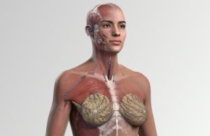
Another company, BioDigital, Inc. was established with the goal of allowing people to see inside the body, using interactive 3D technology. The company developed a cloud platform that makes visualizing the human body through an application easy.
Headquartered in New York City, BioDigital’s 3-D body platform offers both male and female anatomy, in both basic and professional grade detail. Aiding to the overall use of the product, each system is fully segmented, labeled and dissectable for easy configuration to meet any educational need. Together, these features help guide learners through disease progression, treatment and body systems with virtual anatomy tours.
The company’s 3-D body technology also helps learners to understand spatial relationships in the human body with interactive anatomy labels. Another benefit of this Virtual Anatomy tool is that the technology lets users customize the look and feel of 3D anatomy models to emphasize anatomical structures or to highlight key function.
Digital Anatomy Latest News
Elsevier Launches Complete Anatomy Full Female Model
Best Free Anatomy Apps for Mac for Medical Students

UpSurgeOn Academy Revolutionizes Neuroanatomy & Neurosurgery Learning

Institutional Discount for World’s Leading 3D Anatomy Platform: Complete Anatomy From 3D4Medical
Subscribe to HealthySimulation.com’s Newsletter For All the Latest!
Sponsored Content: