ApoQlar’s VSI Promises the Future of Surgery with Hololens
Surgery is a major hub for medical simulation innovation, and apoQlar continues that trend with their Virtual Surgery Intelligence (VSI) line of products which can be used during procedures, for education, and for patient teaching. This German-based startup seeks to move the world, and focuses on creative and active ways for healthcare professionals to learn.
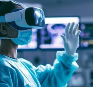
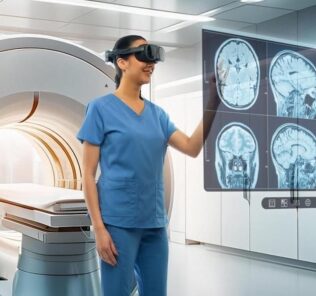
VSI is a smart solution using artificial intelligence which displays MRI and CT images in 3D inside a Mixed/Augmented Reality headset. These images are generated from the original CT or MRI scans. The VSI creates a comprehensive representation, including all anatomical structures, which can be moved around effortlessly in the user’s field of vision. Using anatomical reference points, the VSI recognizes the individual patient and fixes the 3D scans permanently to the surgical site. The surgeon can then remove superfluous layers and structures and adapt the 3D images simultaneously, by using gesture and speech commands.
VSI Products
Sponsored Content:
- VSI Surgery can be used before, during and after the surgery. It helps you prepare yourself before and improves orientation and performance while operating.
- VSI Patient Education can be utilized to explain the progression of the disease to your patients and to educate them about their surgery.
- VSI Education can be applied as a training and education of assistant physicians and young surgeons.
VSI Applications
Neurosurgery: Neurosurgery is concerned with the nervous system focusing on the brain. An advanced medical instrument ‘peels’ the MRI images of the patient’s head to visualize only the required layers of the brain.
ENT: For surgeries in the ENT area, e.g. sinus surgery, a high-quality microscope is important. To ‘slide’ through the 3D MRI images and cut the redundant parts, is another indispensable functionality.
Sponsored Content:
Spinal Surgery: Spinal surgeries afford a high level of precision. Every structure is important. A very accurate tool is needed to help the surgent orientate and operate in-between the finest structures.
Epilepsy: Abnormal patterns of brain waves and brain structures which indicate epilepsy can only be recognized on the MRI images. A clear advantage is provided by the overlap of the 3D MRI images with the surgical site.
Radiology: The 3D view of MRI and CT scans is an excellent tool for radiologists to diagnose a patient’s disease. The functionality to mark structures inside the images is of great value and helpful for further treatment.
New field? Your medical department is not mentioned here? Contact us and become a part of our advisory board in your area of expertise!
Learn more on the VSI website today!
Lance Baily, BA, EMT-B, is the Founder / CEO of HealthySimulation.com, which he started in 2010 while serving as the Director of the Nevada System of Higher Education’s Clinical Simulation Center of Las Vegas. Lance also founded SimGHOSTS.org, the world’s only non-profit organization dedicated to supporting professionals operating healthcare simulation technologies. His co-edited Book: “Comprehensive Healthcare Simulation: Operations, Technology, and Innovative Practice” is cited as a key source for professional certification in the industry. Lance’s background also includes serving as a Simulation Technology Specialist for the LA Community College District, EMS fire fighting, Hollywood movie production, rescue diving, and global travel. He and his wife live with their two brilliant daughters and one crazy dachshund in Las Vegas, Nevada.
Sponsored Content:


