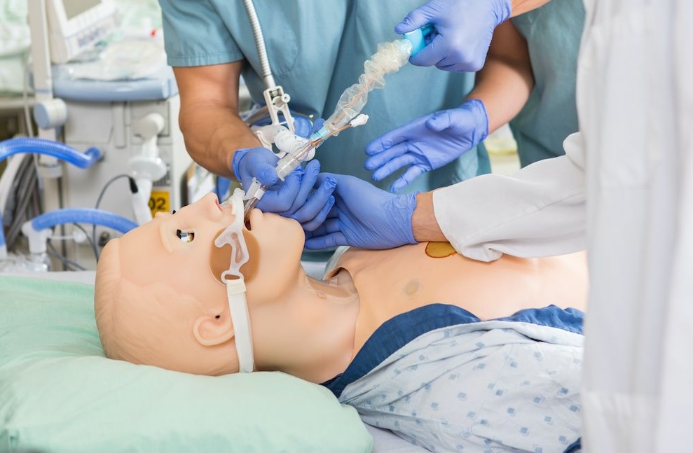
The more closely healthcare simulation matches real life situations (aka fidelity), the more likely that learners will become involved in the learning experience and be less confused. In the clinical setting, healthcare practitioners review past labs or order new ones to help make decisions about appropriate patient care. Lab results should be included in healthcare simulation scenarios where appropriate. Adding lab results provides learners with potentially missing pieces of data that would normally be available. Medical Simulation technologists often come from technical backgrounds with limited knowledge of physiology. By increasing basic physiological knowledge, simulation technicians can add to their understanding of medicine and become more fully involved in simulation operations, research and development.
Today’s article was guest authored by Kim Baily PhD, MSN, RN, CNE, previous Simulation Coordinator for Los Angeles Harbor College and Director of Nursing for El Camino College. Over the past 16 years Kim has developed and implemented several college simulation programs and previously chaired the Southern California Simulation Collaborative. Here, in Part 8 of the Physiology: The Force Behind Healthcare Simulation – A Guide for Techs, Dr. Baily specifically covers ABGs, which stands for Arterial Blood Gases. The entire series is linked below this article.
Warning! If you have not learned about ABGs before, this article may seem complicated but including ABGs in clinical simulation adds to the learning experience! The first part of this article provides a basic introduction to the physiology of ABGs and the second part explains how to interpret ABGs.
Physiology Behind ABG Use
In the intensive care units (ICU) and emergency room settings, the oxygenation status, ventilation and acid-base balance of patients with severe sepsis, acute respiratory failure, and Acute Respiratory Distress Syndrome (ARDS) may be measured from a sample of arterial blood gas.
Arterial Blood Gases are measured from a sample of arterial blood which is collected into an airtight, heparinized syringe and are then transported to the analyzer packed in ice.
A note is made of whether the patient is on oxygen therapy since this will affect interpretation of the ABG. The ABG provides several different components of oxygen and acid/base status:
Oxygenation Status
- Partial pressure of oxygen in blood (PaO2). Normal PaO2 values = 80-100 mmHg
- Estimated normal PaO2 = 100 mmHg – (0.3) age in years
- Hypoxemia (low oxygen level in blood) = PaO2 < 50 mmHg
- Oxygen saturation (SaO2) may be calculated from an ABG unless a co-oximetry is obtained, in which case it is measured i.e. the patient has a pulse oximeter in place.
- An adequate supply of oxygen is vital for normal cellular function. Without circulating oxygen, the body becomes hypoxic and the brain and other cells of the body quickly shut down.
- Note: blood gases can be collected from veins or capillaries however normal oxygen values will be lower.
pH Status
- pH provides a measure of the acidity or alkalinity of the blood.
- Normal values 7.35-7.45. Any number above or below this range is abnormal.
- pH measures the amount of hydrogen ions (H+) by an inverse logarithmic function i.e. the higher the concentration of H+ ions, the lower the pH, the more acidic the body. Think H+ equals acid.
- Acidemia: pH < 7.35
- Conversely, the less H+ ions, the more alkaline and the higher the pH.
- Alkalemia: pH values >7.45
Carbon Dioxide Level
- Partial pressure of carbon dioxide (PaCO2). Normal value = (35-45 mmHg)
- Memory hint – pH 7.35 – 7.45, PaCO2 35-45 mmHg
- Buffers in the circulation act like sponges to absorb excess H+ ions or give off H+ ions when H+ ions are low. The main buffer system is blood is the bicarbonate-carbonic acid buffer system.
- H2O + CO2 < -> H+CO+ < -> [HCO3-] + H+
- As CO2 levels increase in blood, CO2 reacts with water to produce more H+ ions. This leads to more acid and a lower pH. The converse is also true.
- As PaCO2 climbs above 45 mmHg, H+ increases and pH goes down (unless the body compensates).
- As the PaCO2 falls below 35 mmHg, H+ decreases and pH goes up (unless the body compensates).
- Carbon dioxide is blown off from the lungs when we exhale. The faster we breathe, the more CO2 is lost. Conversely, the slower we breathe, the more CO2 will be retained.
- The body responds to changes in pH by either increasing or decreasing respiratory rate to either eliminate or retain CO2.
- Decrease pH (acidosis) → hyperventilation → above equation moves to the left, H+ is reduced → pH returns to normal values.
- Increase in pH (alkalosis) → reduced ventilation → CO2 retained. H+ increases, pH returns to normal values.
- Respiratory acidosis/alkalosis – In an ABG, changes in pH that match changes in PaCO2 are due to a respiratory component.
- Respiratory Acidosis – PaCO2 matches pH change i.e. high PaCO2 → increased acidity (lower pH)
- ABG pH acid < 7.35, PaCO2 > 45 mmHg
- Causes of Respiratory Acidosis:
- Airway obstruction
- COPD
- asthma
- other obstructive lung disease
- CNS depression e.g. by medications
- Sleep disordered breathing
- Neuromuscular impairment – paralysis
- Ventilatory restriction
- Airway obstruction
- Causes of Respiratory Acidosis:
- ABG pH acid < 7.35, PaCO2 > 45 mmHg
- Respiratory Acidosis – PaCO2 matches pH change i.e. high PaCO2 → increased acidity (lower pH)
- Increased CO2 production: shivering, rigors, seizures, malignant hyperthermia, hypermetabolism, increased intake of carbohydrates
- Incorrect mechanical ventilation settings
-
- Respiratory Alkalosis low PaCO2 → increased alkalinity (higher pH).
- ABG pH > 7.45 and the PaCO2 < 35 mmHg
- Causes of Respiratory Alkalosis:
- CNS stimulation: fever, pain, fear, anxiety, CVA, cerebral edema, brain trauma, brain tumor, CNS infection.
- Hypoxemia or hypoxia: lung disease, profound anemia, low FiO2.
- Stimulation of chest receptors: pulmonary edema, pleural effusion, pneumonia, pneumothorax, pulmonary embolus.
- Drugs, hormones: salicylates, catecholamines, medroxyprogesterone, progestins.
- Pregnancy, liver disease, sepsis, hyperthyroidism
- Incorrect mechanical ventilation settings.
- Causes of Respiratory Alkalosis:
- ABG pH > 7.45 and the PaCO2 < 35 mmHg
- Respiratory Alkalosis low PaCO2 → increased alkalinity (higher pH).
Bicarbonate Levels HCO3
The main function of kidneys is retaining or excreting bicarbonate ions (HCO3). Bicarbonate ions neutralizes excess acid in the blood. The prime buffer system is the system of carbonic acid and bicarbonate. Bicarbonate will neutralize the correct numbers of carbonic acid molecules to maintain the correct ratio of 20:1 acid molecules. This 20:1 ratio will preserve the blood pH at the normal range of 7.35 to 7.45. Bicarbonate ions and carbonic acid are constantly being produced and combined in order to keep the optimal pH.
Metabolic acidosis/alkalosis. Metabolic system abnormalities affect acid-balance as acute metabolic acidosis and alkalosis result in acidemia and alkalemia, respectively. Normal HCO3 (22-26 mEq/L)
- Metabolic Acidosis – HCO3 matches pH change i.e. low HCO3 → not enough HCO3 to buffer H+ → increased acidity (lower pH)
- ABG pH acid < 7.35, HCO3 <22 mEq/L
- Causes of Metabolic Acidosis:
- Diabetic ketoacidosis
- Alcoholic and starvation ketoacidosis
- Septic shock
- Renal failure
- Drug or toxin ingestion e.g. methanol intoxication, Isoniazid, Salicylate.
- Gastrointestinal or renal HCO3 loss.
- Causes of Metabolic Acidosis:
- ABG pH acid < 7.35, HCO3 <22 mEq/L
- Metabolic Alkalosis high HCO3 → increased alkalinity (higher pH).
- ABG pH > 7.45 and HCO3 >26 mEq/L
- Causes of Metabolic Alkalosis:
- Kidney disease – renal loss H+
- Heart failure, cirrhosis, nephrotic syndrome, excess ACTH, exogenous steroids.
- Electrolyte imbalances
- GI loss of H+
- Prolonged vomiting, gastric suctioning, villous adenoma, diarrhea
- Hypovolemia
- Diuretic use – loop and thiazide
- Hypokalemia.
- Hypovolemia with Cl- depletion
- Bicarbonate administration
- Kidney disease – renal loss H+
- Causes of Metabolic Alkalosis:
- ABG pH > 7.45 and HCO3 >26 mEq/L
How to Read ABGs
Example 1: pH 7.2, PaCO2 75 mmHg, HCO3 30 mEq/L
- Write down normal values
- pH 7.35-7.454
- PaCO2 35-45 mmHg
- HCO3 22-26 mEq/L
- PaO2 80-100 mmHg
- Determine if pH acidic or alkaline.
- Ex.1. pH acidic <7.35
- Determine whether PaCO2 or HCO3 consistent with pH
- PaCO2 elevated – consistent with acidic pH
- HCO3 elevated – not consistent with acidic pH
- State Cause of acidosis/alkalosis – Respiratory Acidosis
- Relate ABG to clinical situation.
- Determine if body attempting to compensate and whether partial or complete compensation.
- The body is attempting to compensate by increasing HCO3 which is outside the normal range however, even though HCO3 is elevated, the increase in HCO3 is not sufficient to bring the pH back to a normal range. There is only partial compensation.
- Respiratory Acidosis with partial compensation
Example 2: pH 7.36, PaCO2 75 mmHg, HCO3 30 mmHg
- Write down normal values
- pH 7.35-7.454
- PaCO2 35-45 mmHg
- HCO3 22-26 mEq/L
- PaO2 80-100 mmHg
- Determine if pH acidic or alkaline.
- Ex.2 pH normal but towards the acidic end 7.36
- PaCO2 and HCO3 not within normal range, therefore abnormal ABG.
- Determine whether PaCO2 or HCO3 consistent with pH
- PaCO2 elevated – consistent with acidic pH
- HCO3 elevated – not consistent with acidic pH
- State Cause of acidosis/alkalosis – Respiratory Acidosis
- Determine if body attempting to compensate and whether partial or complete compensation.
- The body is attempting to compensate by increasing HCO3 which is outside the normal range and the elevation in HCO3 is sufficient to bring the pH back to the normal range.
- Respiratory acidosis with complete compensation.
- Relate ABG to clinical situation.
Example 3: pH 7.48, PaCO2 50 mmHg, HCO3 30 mmHg
- Write down normal values
- pH 7.35-7.454
- PaCO2 35-45 mmHg
- HCO3 22-26 mEq/L
- PaO2 80-100 mmHg
- Determine if pH acidic or alkaline.
- Ex.3 pH > 7.45 → alkalosis
- PaCO2 and HCO3 not within normal range.
- Determine whether PaCO2 or HCO3 consistent with pH
- PaCO2 elevated – not consistent with alkaline pH
- HCO3 elevated – consistent with alkaline pH
- State Cause of acidosis/alkalosis – Metabolic Alkalosis
- Determine if body attempting to compensate and whether partial or complete compensation.
- The body is attempting to compensate by increasing PaCO2 which is above the normal range however, even though PaCO2 is elevated, the increase in PaCO2 is not sufficient to bring the pH back to a normal range. There is only partial compensation.
- Metabolic Alkalosis with partial compensation.
- Relate ABG to clinical situation.
MIxed Acidosis and Alkalosis – sometimes there may be both respiratory and metabolic acidosis or alkalosis.
- Respiratory and Metabolic Acidosis
- ↓in pH, ↓ in HCO3, ↑ in PaCO2
- Causes: Cardiac arrest, Intoxications, Multi-organ failure
- Respiratory alkalosis with metabolic alkalosis
- ↑in pH, ↑ in HCO3- , ↓ in PaCO2
- Causes: Cirrhosis with diuretics, Pregnancy with vomiting, Over ventilation of COPD
- Other mixed acidosis/alkalosis combinations may be found in complex clinical situations.
ABG Exercises
Test your knowledge, interpret the following ABGs stating if there is full or partial compensation. Answers are at the end of the article.
- pH 7.32, PaCO2 30 mmHg, HCO3 20 mEq/L
- pH 7.32 PaCO2 63 mmHg, HCO3 29 mEq/L
- pH 7.44 PaCO2 55 mmHg, HCO3 30 mEq/L
- pH 7.32 PaCO2 65 mmHg, HCO3 19 mEq/L
Understanding the physiology behind lab results helps simulation technologists become immersed in medical simulation. Inclusion of labs in simulation scenarios increases fidelity and helps learners fully engage in simulation learning experience.
Answers:
- Metabolic acidosis with partial compensation.
- Respiratory acidosis with partial compensation.
- Metabolic alkalosis with complete compensation.
- Combined respiratory and metabolic acidosis.
Read the Entire Physiology: The “Force” Behind Healthcare Simulation HealthySimulation.com Article Series:
- Part 1: Blood Pressure
- Part 2: Heart & Respiratory Rate
- Part 3: Pulse Oximetry
- Part 4: Diabetes
- Part 4B: Hypoglycemia & Excel Template for Simulated EHR
- Part 4C: Insulin
- Part 5: Sepsis
- Part 6A: Hypovolemia (Intro)
- Part 6B: Hypovolemia (Treatment & Simulation Tips)
- Part 7A: IV Fluids & Bags
- Part 7B: IV Pumps & Site Access
- Part 7C: PCA for Pain
- Part 8: ABGs
- Part 9: Sepsis Labs
Learn More About ABGs!
Today’s article was guest authored by Kim Baily PhD, MSN, RN, CNE, previous Simulation Coordinator for Los Angeles Harbor College and Director of Nursing for El Camino College. Over the past 16 years Kim has developed and implemented several college simulation programs and previously chaired the Southern California Simulation Collaborative.
Have a story to share with the global healthcare simulation community? Submit your simulation news and resources here!


