First Transesophageal Echocardiography Simulator with Real Images Created by UW, AHA & Summit Imaging
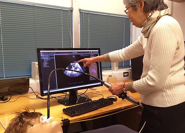
Florence Sheehan, Research Professor at UW’s Division of Cardiology recently shared her work on the DailyUW website regarding a new Cardiac Imaging Technology Simulator, intended to help train and measure image acquisition skills. Transesophageal echocardiography (TEE) technology is not new to the medical field, but Florence Sheehan’s TEE simulator represents a tremendous gain in simulated medical training. The simulator’s development began with a grant from the American Heart Association. Sheehan also credits Larry Nguyen, a UW alum whose Seattle-area company, Summit Imaging, donated time and technical expertise at a pivotal point.
About the New TEE Simulator from UW
Sponsored Content:
Sheehan, a UW research professor and director of the Cardiovascular Research and Training, has succeeded in creating the first TEE simulator that utilizes real patient images. The simulator, which is composed of a mannequin, a diagnostic probe, and a display monitor, allows cardiology fellows and anesthesiology residents of the UW School of Medicine to practice using TEE technology without subjecting patients to the invasive procedure. The simulator also enables the trainees to develop necessary medical skills in a safe environment that allows immediate feedback.
A trainee using Sheehan’s simulator can manipulate either 2-D or 3-D images to obtain the required view of the patient’s heart. Upon achieving a specific view, the simulator can immediately provide the ideal view, as well as information like how far the diagnostic probe was from the ideal position. For Sheehan, the realism of the simulator is what makes it so valuable.
“We’re taking care of real patients,” Sheehan said. “If we train with real images, the fellow will not have to retrain his eye when he is attending to a real patient. If you were learning to drive a car, you could learn by using a simulator, but the simulation would be much improved if you were training with real photos of a road as opposed to drawn images.” The idea for a simulator that incorporated real patient images was born from this desire. In 2014, the American Heart Association awarded Sheehan and her research team the grant to develop such a TEE simulator. Unfortunately, progress on the project soon slowed because the diagnostic probe needed was beyond repair. At this point, Larry Nguyen, the CEO and CTO of Summit Imaging and a Husky alumnus, stepped in to help.
His research team reengineered the probe, making it suitable for use in the simulator. His team also solved several technical problems: his team removed and replaced all the internal components that contained ferrous metals in order to eliminate field noise and prevent interference in the simulated image. The University of Washington team was then able to continue their work developing the image visualization software and reengineering the mannequin’s torso in order to mimic a real TEE operation.
Sponsored Content:
“Real patient images are important because if you only look at an artist’s rendering, you can’t appreciate everything else that an image reveals. For example, the ultrasound modality creates visual artifacts that aren’t meaningful and can be misleading. You have to learn how to distinguish those from an actual pathology,” she said. “The other main consideration is that the image can provide a lot of helpful anatomic information beyond the heart structure you’re focused on. Drawings may not have all that detail.”
Photo Credit: Brian Donohue
Read the full post on DailyUW’s Website!
Lance Baily, BA, EMT-B, is the Founder & CEO of HealthySimulation.com, which he started while serving as the Director of the Nevada System of Higher Education’s Clinical Simulation Center of Las Vegas back in 2010. Lance is also the Founder and acting Advisor to the Board of SimGHOSTS.org, the world’s only non-profit organization dedicated to supporting professionals operating healthcare simulation technologies. His co-edited Book: “Comprehensive Healthcare Simulation: Operations, Technology, and Innovative Practice” is cited as a key source for professional certification in the industry. Lance’s background also includes serving as a Simulation Technology Specialist for the LA Community College District, EMS fire fighting, Hollywood movie production, rescue diving, and global travel. He and his wife Abigail Baily, PhD live in Las Vegas, Nevada with their two amazing daughters.
Sponsored Content:
